A dark long-maned horse standing in mist
The Phoenix Way phoenix emblem

Hoofing Marvellous · Hoof-health framework

The Phoenix Way

Observe the hoof. Follow the horse. Let the evidence lead.

An anatomy-led framework for observing, documenting and supporting the equine foot - built on consistent reference points and the horse’s documented response over time.

A framework for observation

Anatomy first. Nature first. Dogma never.

The Phoenix Way is Hoofing Marvellous’s framework for observing, investigating and supporting the equine foot. It is not a particular trim to copy. It is a way of questioning what we think we know, establishing anatomical reference points and allowing the horse’s documented response over time to guide the next decision.

We do not begin by asking which technique should be applied. We begin by asking what the anatomy, the hoof capsule and the horse are showing us.

A framework - not a formula

It is not

  • A named trim
  • A rigid protocol
  • A product
  • A predetermined answer
  • An intervention that relies on nailed-on shoes or permanently attached fixtures on the horse’s foot

It is

  • A framework for structured observation
  • An anatomy-led way of assessing hoof form
  • A commitment to reproducible documentation
  • A means of testing an interpretation against change over time
  • A basis for proportionate intervention and rehabilitation planning

The same anatomical principles guide the investigation, but the needs of each horse, the condition of each foot and the wider clinical circumstances must still be considered individually.

Question the starting point

What if the diagnosis begins too late in the story?

When a horse presents with pain, hoof distortion or changes on a radiograph, attention often turns immediately to the named condition. Metabolic and systemic illness can be critically important, but the existing mechanical condition of the hoof capsule may receive far less scrutiny.

Heel height, vertical depth, trimming history and the relationship between the hoof capsule and the internal structures all matter. If those factors are not examined, changes seen on an X-ray may be attributed entirely to disease without first asking whether the capsule itself has changed around the foot.

A disease label does not remove the need to assess hoof-capsule balance.

Systemic disease and mechanical hoof-capsule imbalance can coexist, and both require appropriate consideration.

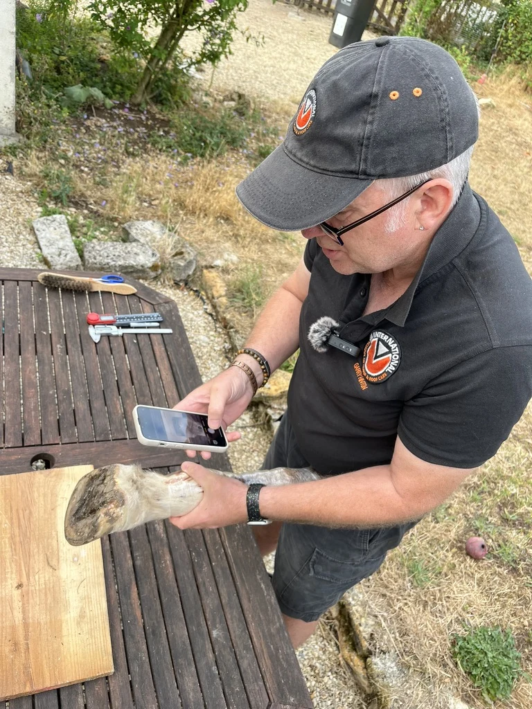
Garry photographing a horse’s foot from above while supporting the lower limb

Observation before intervention

Put down your pitchforks and pick up your cameras.

The invitation is to document before judging. The horse becomes the primary source of evidence: we record what is present before intervention, note what changes and return to the same views after each cycle. A single image can create an impression; a repeatable sequence reveals a pattern.

  1. 01

    Photograph

    Use repeatable views, camera height, distance, lighting and foot placement wherever possible.

  2. 02

    Measure

    Record relevant capsule proportions and anatomical reference points rather than relying on memory or visual impression alone.

  3. 03

    Compare

    Compare the horse with its own previous records - not only with an idealised image of another foot.

  4. 04

    Document

    Record trimming, management, movement, terrain, diet, veterinary findings and other relevant interventions.

  5. 05

    Reassess over time

    Look for direction and pattern across successive cycles. Do not mistake one snapshot for the whole story.

The internal foot as the reference

The capsule must be read in relation to the anatomy within it.

External appearance alone does not tell us whether a hoof capsule is correctly orientated. The Phoenix Way uses consistent anatomical landmarks - including the weight-bearing foot’s True Hard Sole Plane - to assess heel height, vertical depth and the relationship between the capsule and P3.

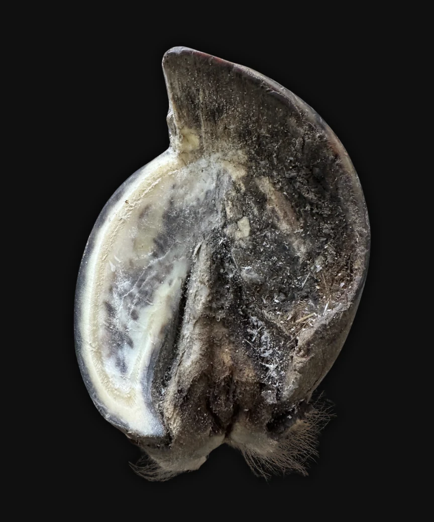
Sectioned equine hoof showing the internal foot and surrounding hoof capsule

A hoof can gradually be trimmed onto the wrong plane. Once that happens, each subsequent trim may follow the existing distortion rather than the anatomy within the foot. Heel height, vertical toe depth and the front-to-back relationship of the capsule can then move further out of proportional balance. The Phoenix Way returns to anatomical reference points so that the foot is not assessed only by its external appearance or by the shape created during previous trimming.

Anatomical constants

Across the Equus genus, equines share a fundamental internal architecture. HM refers to this as the equine blueprint, or Mother Nature’s blueprint. Breed, size and individual form vary, but recurring anatomical landmarks and structural relationships remain. These constants give us dependable reference points for assessing the foot and maintaining proportional balance.

The True Hard Sole Plane

The True Hard Sole Plane largely follows the shape of the solar surface of P3. It gives us a reproducible anatomical reference for orientating the weight-bearing foot. Working in relation to this plane helps us assess heel height, preserve appropriate vertical depth and maintain proportional balance from the front to the back of the hoof capsule.

Biological balance

Balance is judged by what supports viable structure and function - not simply by what appears visually symmetrical on the trimming stand.

Sequential response

An interpretation must remain open to revision when the documented response of the horse does not support it.

Capsule orientation and P3

What changes when the foot moves out of proportional balance?

One conventional explanation describes P3 as rotating or descending independently within the hoof capsule following lamellar failure. Another describes the hoof capsule as rotating around P3. HM’s sequential observations lead us to examine a broader process.

As heel height, vertical toe depth and the front-to-back proportions of the hoof change, the entire foot moves away from proportional balance. The orientation of the capsule, P3 and the ground plane change in relation to one another, while the tissues within the foot are subjected to increasing stress and strain. The radiographic presentation commonly described as “rotation” cannot therefore be understood by looking at P3 or the hoof wall in isolation.

This is why a radiograph must be considered alongside the external capsule, heel height, vertical depth, trimming history, ground plane, positioning and sequential change. A single image cannot show which relationship changed first or how the foot arrived there.

The question is not simply whether P3 moved or the capsule rotated. The question is how the whole foot moved out of proportional balance.

As heel height and front-to-back capsule proportions change, the relationship between P3, the hoof wall and the ground plane changes with them. This sequence illustrates why the entire foot must be assessed rather than describing one structure in isolation.

Intervention changes the system

The toe is not simply excess wall to remove.

Stretch or distortion at the toe should not automatically lead to aggressive removal. Before deciding what to remove, we must understand the anatomical plane, the available vertical depth, the condition of the internal structures and the biological feedback created by ground contact.

Melanie rehabbed her mare who had been suffering from Hoof Capsule Divergence for years, diagnosed as laminitis.

Ground contact and biological feedback

When the toe wall is repeatedly removed from functional ground contact, growth at the front of the capsule can slow relative to growth at the heels. The front and back of the hoof then grow at different relative rates, allowing the capsule to move further away from proportional balance.

A distorted toe is not simply a long toe

What is often described as a long toe may be a distorted toe. When proportional balance is restored and abnormal strain is reduced, a new healing angle can grow down from the coronary band. As this better-connected growth progresses, the older distortion moves down and out of the capsule. In many rehabilitation cases, the distortion can therefore be grown out rather than repeatedly removed from the front.

Across HM’s documented rehabilitation cases, healing angles have grown down and lamellar connection has improved after the hoof capsule was brought into better proportional balance, including cases where the horse’s diet, management and environment remained unchanged. These observations require us to examine the mechanical strain within the capsule rather than assuming that every instance of lamellar distortion has been created by metabolic disease alone.

When the damage limits reconnection

Not every distorted toe can fully reconnect. Where P3 has sustained serious damage, including osteonecrosis, the laminae may be unable to form and maintain the same attachment around the affected bone. In these cases, some degree of toe distortion may remain as a long-term feature of that foot.

The decision is not simply whether to leave or remove the toe. The decision is how to preserve viable structure, restore proportional balance and support the biological feedback needed for healthier growth.

One image is not a trajectory

Reading change through a sequence

These radiographs allow us to follow the relationship between P3 and the changing hoof capsule across successive cycles. The purpose is not to select one image and declare an outcome. It is to examine the direction of change through the complete available sequence.

Chronological sequence · swipe or use controls

August, October and December 2025
February, April, May and September 2026

What the sequence is showing

As proportional balance is restored and maintained, new growth comes down from the coronary band with less distortion. The healing angle progresses down the hoof capsule while the older, stretched growth moves towards the ground and is gradually grown out.

The palmar angle may fluctuate slightly between cycles. HM observes a zone of tolerance in which small fluctuations can occur while the complete capsule remains within proportional front-to-back balance. A single measurement should therefore not be interpreted separately from heel height, vertical depth, the external capsule and the direction of change across the sequence.

By September 2026, the sequence shows how much of the previous toe distortion has progressed down and out of the capsule. The images should be read as a continuing record of growth, balance and internal-external relationships rather than as isolated X-rays.

Radiographs must be interpreted alongside clinical examination, external hoof documentation, positioning and relevant case information.

Core commitments

Five commitments we do not compromise on

  1. 01

    Begin with anatomy

    Use internal structures and viable tissue as the foundation for assessment.

  2. 02

    Observe before intervening

    Establish what is present before deciding what should change.

  3. 03

    Document consistently

    Photograph, measure and record in ways that allow meaningful comparison.

  4. 04

    Make proportionate decisions

    Do no more than the anatomy and current condition of the foot support.

  5. 05

    Let the horse’s response lead

    Reassess every cycle and revise the interpretation when the evidence requires it.

From observation to action

The next decision is earned by the evidence.

  1. 01

    Observe the whole horse

    Consider stance, movement, history, environment, management and all four feet.

  2. 02

    Establish the anatomical references

    Assess the capsule in relation to the internal foot and viable structures.

  3. 03

    Document the starting point

    Create repeatable records before changing the foot.

  4. 04

    Intervene proportionately

    Make only the changes supported by the anatomy and current condition.

  5. 05

    Reassess the response

    Compare successive cycles and allow the horse’s documented response to guide what happens next.

This is not passive observation. It is disciplined clinical reasoning: act, document, reassess and remain willing to change the interpretation.

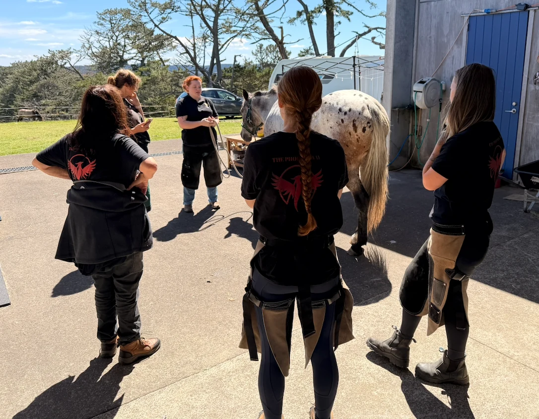
Hoofing Marvellous team members observing a horse together outdoors

The horse in front of us

Rehabilitation, long-term management or palliative care

The Phoenix Way begins with the horse and the foot that exist today, not the foot we wish were there. Some horses have sufficient viable structure to make meaningful rehabilitation possible. Others have permanent changes that limit how much recovery the foot can achieve. Extensive osteonecrosis, severe caudal heel failure, advanced arthritis and other irreversible structural damage may prevent full rehabilitation.

Some horses can still be supported comfortably through careful long-term management or palliative care, sometimes for many years. For others, the remaining damage may be too extensive for hoof rehabilitation to offer a reasonable or lasting improvement. Professional responsibility means recognising the difference. An HMB Professional should remain calm, honest and kind, helping the owner understand whether the realistic pathway is rehabilitation, continuing management, palliative support or an acknowledgement that the foot can no longer be helped through hoof care alone.

The aim is not to promise recovery. It is to understand what remains possible and make decisions in the best interests of the horse.

Where do you go from here?

Explore The Phoenix Way

Whether you need help with your own horse or want to understand the anatomy and clinical thinking in greater depth, choose the pathway that fits where you are now.

I need support for my horse

Request in-person or online support and allow the HM team to explore the available options.

Find Professional Support

I want to understand more

Learn to observe the hoof and the horse with greater confidence through Phoenix Academy.

Explore Phoenix Academy

I want practical learning

Join the HM team for 3 days of anatomy-led teaching, cadaver work, demonstrations and practical learning.

View Workshops
The Phoenix Way Facebook banner: the red phoenix emblem with the words The Phoenix Way and the tagline Where hoof health takes flight, beside a black-and-white portrait of a dark horse.

I want to join the free Phoenix Way community

Join more than 15,000 members in The Phoenix Way: Path 2 Hoof Health, our free Facebook group where owners and professionals share rehabilitation journeys, documented cases, questions and ongoing learning.

Join the Free Facebook Group